3w1y

X-ray diffraction
2.3Å resolution

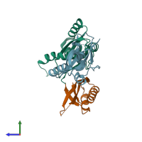
Crystal structure of T brucei ATG8.2 in complex with E coli S10

Released:
Entry authors: Yuan YA, Wang C

Function and Biology Details

Structure analysis Details

Assembly composition:
hetero trimer (preferred)
PDBe Complex ID:
PDB-CPX-141622 (preferred)
Entry contents:
2 distinct polypeptide molecules
Macromolecules (2 distinct):
Autophagy-related protein Chains: A, B
Molecule details ›
Chains: A, B
Length: 137 amino acids
Theoretical weight: 15.63 KDa
Source organism: Trypanosoma brucei brucei TREU927
Expression system: Escherichia coli
UniProt:
  • Canonical: Q57WE7 (Residues: 1-134; Coverage: 100%)
Gene names: Tb07.28B13.800, Tb927.7.3320
Sequence domains: Autophagy protein Atg8 ubiquitin like
Structure domains: Phosphatidylinositol 3-kinase Catalytic Subunit; Chain A, domain 1
Small ribosomal subunit protein uS10 Chain: C
Molecule details ›
Chain: C
Length: 103 amino acids
Theoretical weight: 11.76 KDa
Source organism: Escherichia coli K-12
Expression system: Escherichia coli
UniProt:
  • Canonical: P0A7R5 (Residues: 1-103; Coverage: 100%)
Gene names: JW3283, b3321, nusE, rpsJ
Sequence domains: Ribosomal protein S10p/S20e
Structure domains: Ribosomal protein S10 domain

Ligands and Environments

No bound ligands
No modified residues

Experiments and Validation Details

Entry percentile scores
X-ray source: NSLS BEAMLINE X29A
Spacegroup: P21
Unit cell:
a: 48.012Å b: 61.197Å c: 67.636Å
α: 90° β: 110.77° γ: 90°
R-values:
R R work R free
0.214 0.203 0.256
Expression system: Escherichia coli