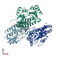
3ujp

X-ray diffraction
2.7Å resolution

Structure of MntC protein at 2.7A

Released:
Source organism: Synechocystis sp. PCC 6803
Primary publication:
Arginine 116 stabilizes the entrance to the metal ion-binding site of the MntC protein.
Acta Crystallogr Sect F Struct Biol Cryst Commun 69 237-42 (2013)
PMID: 23519795

Function and Biology Details

Biochemical function:
Biological process:
Cellular component:
  • not assigned

Structure analysis Details

Assembly composition:
homo hexamer (preferred)
Assembly name:
PDBe Complex ID:
PDB-CPX-181256 (preferred)
Entry contents:
1 distinct polypeptide molecule
Macromolecule:
Mn transporter MntC Chains: A, B, C
Molecule details ›
Chains: A, B, C
Length: 307 amino acids
Theoretical weight: 33.78 KDa
Source organism: Synechocystis sp. PCC 6803
Expression system: Escherichia coli BL21
UniProt:
  • Canonical: Q79EF9 (Residues: 24-330; Coverage: 99%)
Gene name: mntC
Sequence domains: Zinc-uptake complex component A periplasmic
Structure domains: Nitrogenase molybdenum iron protein domain

Ligands and Environments

3 bound ligands:
No modified residues

Experiments and Validation Details

Entry percentile scores
X-ray source: ESRF BEAMLINE ID23-1
Spacegroup: P3121
Unit cell:
a: 127.52Å b: 127.52Å c: 89.73Å
α: 90° β: 90° γ: 120°
R-values:
R R work R free
0.233 0.231 0.284
Expression system: Escherichia coli BL21