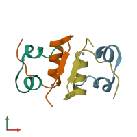
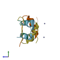
3ins

Hybrid

STRUCTURE OF INSULIN. RESULTS OF JOINT NEUTRON AND X-RAY REFINEMENT

Released:
Source organism: Sus scrofa
Primary publication:
Structure of insulin: results of joint neutron and X-ray refinement.
Acta Crystallogr B 45 ( Pt 1) 99-107 (1989)
PMID: 2695122

Function and Biology Details

Biochemical function:
  • not assigned
Biological process:
  • not assigned
Cellular component:
  • not assigned

Structure analysis Details

Assemblies composition:
hetero dimer (preferred)
hetero dodecamer
Assembly name:
PDBe Complex ID:
PDB-CPX-134237 (preferred)
Entry contents:
2 distinct polypeptide molecules
Macromolecules (2 distinct):
Insulin A chain Chains: A, C
Molecule details ›
Chains: A, C
Length: 21 amino acids
Theoretical weight: 2.38 KDa
Source organism: Sus scrofa
Expression system: Not provided
UniProt:
  • Canonical: P01315 (Residues: 88-108; Coverage: 25%)
Gene name: INS
Insulin B chain Chains: B, D
Molecule details ›
Chains: B, D
Length: 30 amino acids
Theoretical weight: 3.4 KDa
Source organism: Sus scrofa
Expression system: Not provided
UniProt:
  • Canonical: P01315 (Residues: 25-54; Coverage: 36%)
Gene name: INS
Sequence domains: Insulin/IGF/Relaxin family

Ligands and Environments

2 bound ligands:
No modified residues

Experiments and Validation Details

Experimental Method: X-ray diffraction, Neutron Diffraction
Spacegroup: R3
Unit cell:
a: 82.5Å b: 82.5Å c: 34Å
α: 90° β: 90° γ: 120°
R-values:
R R work R free
0.182 0.182 not available
Expression system: Not provided