Function and Biology Details
Reactions catalysed:
S-adenosyl-L-methionine + a 5'-(5'-triphosphoguanosine)-[mRNA] = S-adenosyl-L-homocysteine + a 5'-(N(7)-methyl 5'-triphosphoguanosine)-[mRNA]
S-adenosyl-L-methionine + a 5'-(N(7)-methyl 5'-triphosphoguanosine)-(ribonucleotide)-[mRNA] = S-adenosyl-L-homocysteine + a 5'-(N(7)-methyl 5'-triphosphoguanosine)-(2'-O-methyl-ribonucleotide)-[mRNA]
Nucleoside triphosphate + RNA(n) = diphosphate + RNA(n+1)
Selective hydrolysis of -Xaa-Xaa-|-Yaa- bonds in which each of the Xaa can be either Arg or Lys and Yaa can be either Ser or Ala.
NTP + H(2)O = NDP + phosphate
ATP + H(2)O = ADP + phosphate
Biochemical function:
Biological process:
Cellular component:
- not assigned
Structure analysis Details
Assembly composition:
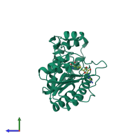
monomeric (preferred)
Assembly name:
Serine protease NS3 (preferred)
PDBe Complex ID:
PDB-CPX-136776 (preferred)
Entry contents:
1 distinct polypeptide molecule
Macromolecule:


