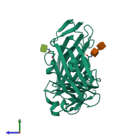
3u74

X-ray diffraction
2.39Å resolution

Crystal structure of stabilized human uPAR mutant

Released:
Source organism: Homo sapiens
Primary publication:
Crystal structure of the urokinase receptor in a ligand-free form.
J Mol Biol 416 629-41 (2012)
PMID: 22285761

Function and Biology Details

Structure analysis Details

Assembly composition:
monomeric (preferred)
PDBe Complex ID:
PDB-CPX-169884 (preferred)
Entry contents:
1 distinct polypeptide molecule
Macromolecules (2 distinct):
Urokinase plasminogen activator surface receptor Chain: U
Molecule details ›
Chain: U
Length: 283 amino acids
Theoretical weight: 31.46 KDa
Source organism: Homo sapiens
Expression system: Drosophila melanogaster
UniProt:
  • Canonical: Q03405 (Residues: 23-305; Coverage: 90%)
Gene names: MO3, PLAUR, UPAR
Sequence domains: u-PAR/Ly-6 domain
Structure domains: CD59

Ligands and Environments

Carbohydrate polymer : NEW Components: NAG
1 bound ligand:
No modified residues

Experiments and Validation Details

Entry percentile scores
X-ray source: NSLS BEAMLINE X29A
Spacegroup: R3
Unit cell:
a: 111.235Å b: 111.235Å c: 78.571Å
α: 90° β: 90° γ: 120°
R-values:
R R work R free
0.214 0.213 0.252
Expression system: Drosophila melanogaster