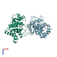
3rpt

X-ray diffraction
1.3Å resolution

Crystal structure of the anti-HIV b12 scaffold protein

Released:

Function and Biology Details

Reaction catalysed:
Endohydrolysis of (1->4)-beta-D-glucosidic linkages in cellulose, lichenin and cereal beta-D-glucans
Biological process:
Cellular component:
  • not assigned

Structure analysis Details

Assembly composition:
monomeric (preferred)
Assembly name:
PDBe Complex ID:
PDB-CPX-150674 (preferred)
Entry contents:
1 distinct polypeptide molecule
Macromolecule:
Endoglucanase E-2 Chains: A, X
Molecule details ›
Chains: A, X
Length: 282 amino acids
Theoretical weight: 29.83 KDa
Source organism: Thermobifida fusca
Expression system: Escherichia coli
UniProt:
  • Canonical: P26222 (Residues: 33-317; Coverage: 67%)
Gene name: celB
Sequence domains: Glycosyl hydrolases family 6
Structure domains: 1, 4-beta cellobiohydrolase

Ligands and Environments

1 bound ligand:
No modified residues

Experiments and Validation Details

Entry percentile scores
X-ray source: APS BEAMLINE 22-ID
Spacegroup: P1
Unit cell:
a: 45.867Å b: 46.24Å c: 62.282Å
α: 78.57° β: 81.8° γ: 61.11°
R-values:
R R work R free
0.175 0.174 0.202
Expression system: Escherichia coli