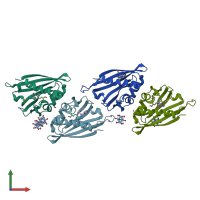
3c0v

X-ray diffraction
1.8Å resolution

Crystal structure of cytokinin-specific binding protein in complex with cytokinin and Ta6Br12

Released:
Source organism: Vigna radiata
Primary publication:
MAD phasing using the (Ta6Br12)2+ cluster: a retrospective study.
Acta Crystallogr D Biol Crystallogr 64 595-606 (2008)
PMID: 18453695

Function and Biology Details

Structure analysis Details

Assemblies composition:
monomeric
homo dimer (preferred)
PDBe Complex ID:
PDB-CPX-103684 (preferred)
Entry contents:
1 distinct polypeptide molecule
Macromolecule:
Phytohormone-binding protein CSBP Chains: A, B, C, D
Molecule details ›
Chains: A, B, C, D
Length: 155 amino acids
Theoretical weight: 17.61 KDa
Source organism: Vigna radiata
Expression system: Escherichia coli BL21
UniProt:
  • Canonical: A0A1S3THR8 (Residues: 1-155; Coverage: 100%)
Gene names: CSBP, LOC106755646
Sequence domains: Pathogenesis-related protein Bet v 1 family
Structure domains: Alpha-D-Glucose-1,6-Bisphosphate; Chain A, domain 4

Ligands and Environments

No modified residues

Experiments and Validation Details

Entry percentile scores
X-ray source: EMBL/DESY, HAMBURG BEAMLINE BW7A
Spacegroup: P64
Unit cell:
a: 113.219Å b: 113.219Å c: 85.077Å
α: 90° β: 90° γ: 120°
R-values:
R R work R free
0.159 0.158 0.207
Expression system: Escherichia coli BL21