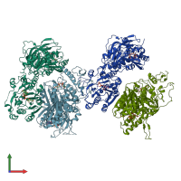
Function and Biology Details
Reactions catalysed:
UDP-N-acetyl-alpha-D-galactosamine + [protein]-3-O-(beta-D-GlcA-(1->3)-(beta-D-GalNAc-(1->4)-beta-D-GlcA-(1->3))(n)-beta-D-GalNAc-(1->4)-beta-D-GlcA-(1->3)-beta-D-Gal-(1->3)-beta-D-Gal-(1->4)-beta-D-Xyl)-L-serine = UDP + [protein]-3-O-((beta-D-GalNAc-(1->4)-beta-D-GlcA-(1->3))(n+1)-beta-D-GalNAc-(1->4)-beta-D-GlcA-(1->3)-beta-D-Gal-(1->3)-beta-D-Gal-(1->4)-beta-D-Xyl)-L-serine
UDP-alpha-D-glucuronate + [protein]-3-O-((beta-D-GalNAc-(1->4)-beta-D-GlcA-(1->3))(n)-beta-D-GalNAc-(1->4)-beta-D-GlcA-(1->3)-beta-D-Gal-(1->3)-beta-D-Gal-(1->4)-beta-D-Xyl)-L-serine = UDP + [protein]-3-O-(beta-D-GlcA-(1->3)-(beta-D-GalNAc-(1->4)-beta-D-GlcA-(1->3))(n)-beta-D-GalNAc-(1->4)-beta-D-GlcA-(1->3)-beta-D-Gal-(1->3)-beta-D-Gal-(1->4)-beta-D-Xyl)-L-serine
Biochemical function:
Biological process:
Cellular component:
- not assigned
Sequence domains:
Structure analysis Details
Assembly composition:
homo dimer (preferred)
Assembly name:
Chondroitin synthase (preferred)
PDBe Complex ID:
PDB-CPX-185230 (preferred)
Entry contents:
1 distinct polypeptide molecule
Macromolecule:


