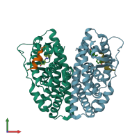
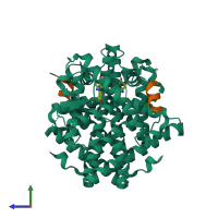
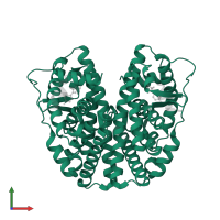
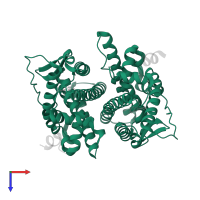
Assemblies
Assembly Name:
Nuclear receptor coactivator 2 and Estrogen receptor
Multimeric state:
hetero tetramer
Accessible surface area:
20597.29 Å2
Buried surface area:
6520.31 Å2
Dissociation area:
1,011.66
Å2
Dissociation energy (ΔGdiss):
6.61
kcal/mol
Dissociation entropy (TΔSdiss):
14.44
kcal/mol
Symmetry number:
2
PDBe Complex ID:
PDB-CPX-136930
Macromolecules
Chains: A, B
Length: 258 amino acids
Theoretical weight: 29.39 KDa
Source organism: Homo sapiens
Expression system: Escherichia coli
UniProt:
Pfam: Ligand-binding domain of nuclear hormone receptor
InterPro:
Length: 258 amino acids
Theoretical weight: 29.39 KDa
Source organism: Homo sapiens
Expression system: Escherichia coli
UniProt:
- Canonical:
 P03372 (Residues: 298-554; Coverage: 43%)
Pfam: Ligand-binding domain of nuclear hormone receptor
InterPro:
- Nuclear hormone receptor-like domain superfamily
- Nuclear hormone receptor, ligand-binding domain
- Nuclear hormone receptor


