Assemblies
Multimeric state:
hetero hexamer
Accessible surface area:
69664.35 Å2
Buried surface area:
32342.84 Å2
Dissociation area:
3,383.85
Å2
Dissociation energy (ΔGdiss):
56.24
kcal/mol
Dissociation entropy (TΔSdiss):
16.68
kcal/mol
Symmetry number:
2
PDBe Complex ID:
PDB-CPX-169747
Multimeric state:
hetero hexamer
Accessible surface area:
69934.22 Å2
Buried surface area:
32287.17 Å2
Dissociation area:
3,381.82
Å2
Dissociation energy (ΔGdiss):
57.4
kcal/mol
Dissociation entropy (TΔSdiss):
16.68
kcal/mol
Symmetry number:
2
PDBe Complex ID:
PDB-CPX-169747
Multimeric state:
hetero hexamer
Accessible surface area:
69868.84 Å2
Buried surface area:
32470.9 Å2
Dissociation area:
5,015.52
Å2
Dissociation energy (ΔGdiss):
57.9
kcal/mol
Dissociation entropy (TΔSdiss):
29.47
kcal/mol
Symmetry number:
2
PDBe Complex ID:
PDB-CPX-169747
Macromolecules
Chains: A, D, G, J, M, P
Length: 445 amino acids
Theoretical weight: 50.16 KDa
Source organism: Cereibacter sphaeroides
Expression system: Cereibacter sphaeroides
UniProt:
Pfam:
InterPro:
Length: 445 amino acids
Theoretical weight: 50.16 KDa
Source organism: Cereibacter sphaeroides
Expression system: Cereibacter sphaeroides
UniProt:
- Canonical:
 Q02761 (Residues: 1-445; Coverage: 100%)
Pfam:
InterPro:
- Cytochrome b/b6-like domain superfamily
- Cytochrome b
- Cytochrome b/b6, N-terminal domain
- Di-haem cytochrome, transmembrane
- Cytochrome b/b6, C-terminal
- Cytochrome b/b6, C-terminal domain superfamily
Chains: B, E, H, K, N, Q
Length: 269 amino acids
Theoretical weight: 29.37 KDa
Source organism: Cereibacter sphaeroides
Expression system: Cereibacter sphaeroides
UniProt:
Pfam: Cytochrome C1 family
InterPro:
CATH:
Length: 269 amino acids
Theoretical weight: 29.37 KDa
Source organism: Cereibacter sphaeroides
Expression system: Cereibacter sphaeroides
UniProt:
- Canonical:
 Q02760 (Residues: 23-285; Coverage: 100%)
Pfam: Cytochrome C1 family
InterPro:
CATH:
Chains: C, F, I, L, O, R
Length: 187 amino acids
Theoretical weight: 19.92 KDa
Source organism: Cereibacter sphaeroides
Expression system: Cereibacter sphaeroides
UniProt:
Pfam:
InterPro:
Length: 187 amino acids
Theoretical weight: 19.92 KDa
Source organism: Cereibacter sphaeroides
Expression system: Cereibacter sphaeroides
UniProt:
- Canonical:
 Q02762 (Residues: 1-187; Coverage: 100%)
Pfam:
InterPro:
- Twin-arginine translocation pathway, signal sequence
- Ubiquitinol-cytochrome C reductase, Fe-S subunit, TAT signal
- Rieske iron-sulphur protein
- Ubiquinol-cytochrome c reductase, iron-sulphur subunit
- Twin-arginine translocation pathway, signal sequence, bacterial/archaeal
- Rieske [2Fe-2S] iron-sulphur domain superfamily
- Rieske [2Fe-2S] iron-sulphur domain
- Rieske iron-sulphur protein, C-terminal


![The deposited structure of PDB entry <span class='highlight'>2fyn</span> coloured by chain, front view. This structure contains: <ul class='image_legend_ul'><li class='image_legend_li'>6 copies of <span class='highlight'>Cytochrome b</span>;</li> <li class='image_legend_li'>6 copies of <span class='highlight'>Cytochrome c1</span>;</li> <li class='image_legend_li'>6 copies of <span class='highlight'>Ubiquinol-cytochrome c reductase iron-sulfur subunit</span>;</li> <li class='image_legend_li'>18 copies of <span class='highlight'>PROTOPORPHYRIN IX CONTAINING FE</span>;</li> <li class='image_legend_li'>6 copies of <span class='highlight'>STIGMATELLIN A</span>;</li> <li class='image_legend_li'>6 copies of <span class='highlight'>(1R)-2-{[(R)-(2-AMINOETHOXY)(HYDROXY)PHOSPHORYL]OXY}-1-[(DODECANOYLOXY)METHYL]ETHYL (9Z)-OCTADEC-9-ENOATE</span>;</li> <li class='image_legend_li'>6 copies of <span class='highlight'>FE2/S2 (INORGANIC) CLUSTER</span>.</li></ul> PDB entry 2fyn coloured by chain, front view.](https://www.ebi.ac.uk/pdbe/static/entry/2fyn_deposited_chain_front_image-200x200.png)
![The deposited structure of PDB entry <span class='highlight'>2fyn</span> coloured by chain, side view. This structure contains: <ul class='image_legend_ul'><li class='image_legend_li'>6 copies of <span class='highlight'>Cytochrome b</span>;</li> <li class='image_legend_li'>6 copies of <span class='highlight'>Cytochrome c1</span>;</li> <li class='image_legend_li'>6 copies of <span class='highlight'>Ubiquinol-cytochrome c reductase iron-sulfur subunit</span>;</li> <li class='image_legend_li'>18 copies of <span class='highlight'>PROTOPORPHYRIN IX CONTAINING FE</span>;</li> <li class='image_legend_li'>6 copies of <span class='highlight'>STIGMATELLIN A</span>;</li> <li class='image_legend_li'>6 copies of <span class='highlight'>(1R)-2-{[(R)-(2-AMINOETHOXY)(HYDROXY)PHOSPHORYL]OXY}-1-[(DODECANOYLOXY)METHYL]ETHYL (9Z)-OCTADEC-9-ENOATE</span>;</li> <li class='image_legend_li'>6 copies of <span class='highlight'>FE2/S2 (INORGANIC) CLUSTER</span>.</li></ul> PDB entry 2fyn coloured by chain, side view.](https://www.ebi.ac.uk/pdbe/static/entry/2fyn_deposited_chain_side_image-200x200.png)
![The deposited structure of PDB entry <span class='highlight'>2fyn</span> coloured by chain, top view. This structure contains: <ul class='image_legend_ul'><li class='image_legend_li'>6 copies of <span class='highlight'>Cytochrome b</span>;</li> <li class='image_legend_li'>6 copies of <span class='highlight'>Cytochrome c1</span>;</li> <li class='image_legend_li'>6 copies of <span class='highlight'>Ubiquinol-cytochrome c reductase iron-sulfur subunit</span>;</li> <li class='image_legend_li'>18 copies of <span class='highlight'>PROTOPORPHYRIN IX CONTAINING FE</span>;</li> <li class='image_legend_li'>6 copies of <span class='highlight'>STIGMATELLIN A</span>;</li> <li class='image_legend_li'>6 copies of <span class='highlight'>(1R)-2-{[(R)-(2-AMINOETHOXY)(HYDROXY)PHOSPHORYL]OXY}-1-[(DODECANOYLOXY)METHYL]ETHYL (9Z)-OCTADEC-9-ENOATE</span>;</li> <li class='image_legend_li'>6 copies of <span class='highlight'>FE2/S2 (INORGANIC) CLUSTER</span>.</li></ul> PDB entry 2fyn coloured by chain, top view.](https://www.ebi.ac.uk/pdbe/static/entry/2fyn_deposited_chain_top_image-200x200.png)
![Hetero hexameric assembly 1 of PDB entry <span class='highlight'>2fyn</span> coloured by chemically distinct molecules, front view. This structure contains: <ul class='image_legend_ul'><li class='image_legend_li'>2 copies of <span class='highlight'>Cytochrome b</span>;</li> <li class='image_legend_li'>2 copies of <span class='highlight'>Cytochrome c1</span>;</li> <li class='image_legend_li'>2 copies of <span class='highlight'>Ubiquinol-cytochrome c reductase iron-sulfur subunit</span>;</li> <li class='image_legend_li'>6 copies of <span class='highlight'>PROTOPORPHYRIN IX CONTAINING FE</span>;</li> <li class='image_legend_li'>2 copies of <span class='highlight'>STIGMATELLIN A</span>;</li> <li class='image_legend_li'>2 copies of <span class='highlight'>(1R)-2-{[(R)-(2-AMINOETHOXY)(HYDROXY)PHOSPHORYL]OXY}-1-[(DODECANOYLOXY)METHYL]ETHYL (9Z)-OCTADEC-9-ENOATE</span>;</li> <li class='image_legend_li'>2 copies of <span class='highlight'>FE2/S2 (INORGANIC) CLUSTER</span>.</li></ul>} Hetero hexameric assembly 1 of PDB entry 2fyn coloured by chemically distinct molecules, front view.](https://www.ebi.ac.uk/pdbe/static/entry/2fyn_assembly_1_chemically_distinct_molecules_front_image-200x200.png)
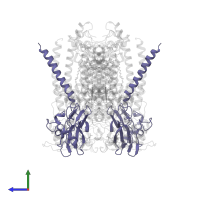
![Hetero hexameric assembly 1 of PDB entry <span class='highlight'>2fyn</span> coloured by chemically distinct molecules, side view. This structure contains: <ul class='image_legend_ul'><li class='image_legend_li'>2 copies of <span class='highlight'>Cytochrome b</span>;</li> <li class='image_legend_li'>2 copies of <span class='highlight'>Cytochrome c1</span>;</li> <li class='image_legend_li'>2 copies of <span class='highlight'>Ubiquinol-cytochrome c reductase iron-sulfur subunit</span>;</li> <li class='image_legend_li'>6 copies of <span class='highlight'>PROTOPORPHYRIN IX CONTAINING FE</span>;</li> <li class='image_legend_li'>2 copies of <span class='highlight'>STIGMATELLIN A</span>;</li> <li class='image_legend_li'>2 copies of <span class='highlight'>(1R)-2-{[(R)-(2-AMINOETHOXY)(HYDROXY)PHOSPHORYL]OXY}-1-[(DODECANOYLOXY)METHYL]ETHYL (9Z)-OCTADEC-9-ENOATE</span>;</li> <li class='image_legend_li'>2 copies of <span class='highlight'>FE2/S2 (INORGANIC) CLUSTER</span>.</li></ul>} Hetero hexameric assembly 1 of PDB entry 2fyn coloured by chemically distinct molecules, side view.](https://www.ebi.ac.uk/pdbe/static/entry/2fyn_assembly_1_chemically_distinct_molecules_side_image-200x200.png)
![Hetero hexameric assembly 1 of PDB entry <span class='highlight'>2fyn</span> coloured by chemically distinct molecules, top view. This structure contains: <ul class='image_legend_ul'><li class='image_legend_li'>2 copies of <span class='highlight'>Cytochrome b</span>;</li> <li class='image_legend_li'>2 copies of <span class='highlight'>Cytochrome c1</span>;</li> <li class='image_legend_li'>2 copies of <span class='highlight'>Ubiquinol-cytochrome c reductase iron-sulfur subunit</span>;</li> <li class='image_legend_li'>6 copies of <span class='highlight'>PROTOPORPHYRIN IX CONTAINING FE</span>;</li> <li class='image_legend_li'>2 copies of <span class='highlight'>STIGMATELLIN A</span>;</li> <li class='image_legend_li'>2 copies of <span class='highlight'>(1R)-2-{[(R)-(2-AMINOETHOXY)(HYDROXY)PHOSPHORYL]OXY}-1-[(DODECANOYLOXY)METHYL]ETHYL (9Z)-OCTADEC-9-ENOATE</span>;</li> <li class='image_legend_li'>2 copies of <span class='highlight'>FE2/S2 (INORGANIC) CLUSTER</span>.</li></ul>} Hetero hexameric assembly 1 of PDB entry 2fyn coloured by chemically distinct molecules, top view.](https://www.ebi.ac.uk/pdbe/static/entry/2fyn_assembly_1_chemically_distinct_molecules_top_image-200x200.png)
![Hetero hexameric assembly 2 of PDB entry <span class='highlight'>2fyn</span> coloured by chemically distinct molecules, front view. This structure contains: <ul class='image_legend_ul'><li class='image_legend_li'>2 copies of <span class='highlight'>Cytochrome b</span>;</li> <li class='image_legend_li'>2 copies of <span class='highlight'>Cytochrome c1</span>;</li> <li class='image_legend_li'>2 copies of <span class='highlight'>Ubiquinol-cytochrome c reductase iron-sulfur subunit</span>;</li> <li class='image_legend_li'>6 copies of <span class='highlight'>PROTOPORPHYRIN IX CONTAINING FE</span>;</li> <li class='image_legend_li'>2 copies of <span class='highlight'>STIGMATELLIN A</span>;</li> <li class='image_legend_li'>2 copies of <span class='highlight'>(1R)-2-{[(R)-(2-AMINOETHOXY)(HYDROXY)PHOSPHORYL]OXY}-1-[(DODECANOYLOXY)METHYL]ETHYL (9Z)-OCTADEC-9-ENOATE</span>;</li> <li class='image_legend_li'>2 copies of <span class='highlight'>FE2/S2 (INORGANIC) CLUSTER</span>.</li></ul>} Hetero hexameric assembly 2 of PDB entry 2fyn coloured by chemically distinct molecules, front view.](https://www.ebi.ac.uk/pdbe/static/entry/2fyn_assembly_2_chemically_distinct_molecules_front_image-200x200.png)
![Hetero hexameric assembly 2 of PDB entry <span class='highlight'>2fyn</span> coloured by chemically distinct molecules, side view. This structure contains: <ul class='image_legend_ul'><li class='image_legend_li'>2 copies of <span class='highlight'>Cytochrome b</span>;</li> <li class='image_legend_li'>2 copies of <span class='highlight'>Cytochrome c1</span>;</li> <li class='image_legend_li'>2 copies of <span class='highlight'>Ubiquinol-cytochrome c reductase iron-sulfur subunit</span>;</li> <li class='image_legend_li'>6 copies of <span class='highlight'>PROTOPORPHYRIN IX CONTAINING FE</span>;</li> <li class='image_legend_li'>2 copies of <span class='highlight'>STIGMATELLIN A</span>;</li> <li class='image_legend_li'>2 copies of <span class='highlight'>(1R)-2-{[(R)-(2-AMINOETHOXY)(HYDROXY)PHOSPHORYL]OXY}-1-[(DODECANOYLOXY)METHYL]ETHYL (9Z)-OCTADEC-9-ENOATE</span>;</li> <li class='image_legend_li'>2 copies of <span class='highlight'>FE2/S2 (INORGANIC) CLUSTER</span>.</li></ul>} Hetero hexameric assembly 2 of PDB entry 2fyn coloured by chemically distinct molecules, side view.](https://www.ebi.ac.uk/pdbe/static/entry/2fyn_assembly_2_chemically_distinct_molecules_side_image-200x200.png)
![Hetero hexameric assembly 2 of PDB entry <span class='highlight'>2fyn</span> coloured by chemically distinct molecules, top view. This structure contains: <ul class='image_legend_ul'><li class='image_legend_li'>2 copies of <span class='highlight'>Cytochrome b</span>;</li> <li class='image_legend_li'>2 copies of <span class='highlight'>Cytochrome c1</span>;</li> <li class='image_legend_li'>2 copies of <span class='highlight'>Ubiquinol-cytochrome c reductase iron-sulfur subunit</span>;</li> <li class='image_legend_li'>6 copies of <span class='highlight'>PROTOPORPHYRIN IX CONTAINING FE</span>;</li> <li class='image_legend_li'>2 copies of <span class='highlight'>STIGMATELLIN A</span>;</li> <li class='image_legend_li'>2 copies of <span class='highlight'>(1R)-2-{[(R)-(2-AMINOETHOXY)(HYDROXY)PHOSPHORYL]OXY}-1-[(DODECANOYLOXY)METHYL]ETHYL (9Z)-OCTADEC-9-ENOATE</span>;</li> <li class='image_legend_li'>2 copies of <span class='highlight'>FE2/S2 (INORGANIC) CLUSTER</span>.</li></ul>} Hetero hexameric assembly 2 of PDB entry 2fyn coloured by chemically distinct molecules, top view.](https://www.ebi.ac.uk/pdbe/static/entry/2fyn_assembly_2_chemically_distinct_molecules_top_image-200x200.png)
![Hetero hexameric assembly 3 of PDB entry <span class='highlight'>2fyn</span> coloured by chemically distinct molecules, front view. This structure contains: <ul class='image_legend_ul'><li class='image_legend_li'>2 copies of <span class='highlight'>Cytochrome b</span>;</li> <li class='image_legend_li'>2 copies of <span class='highlight'>Cytochrome c1</span>;</li> <li class='image_legend_li'>2 copies of <span class='highlight'>Ubiquinol-cytochrome c reductase iron-sulfur subunit</span>;</li> <li class='image_legend_li'>6 copies of <span class='highlight'>PROTOPORPHYRIN IX CONTAINING FE</span>;</li> <li class='image_legend_li'>2 copies of <span class='highlight'>STIGMATELLIN A</span>;</li> <li class='image_legend_li'>2 copies of <span class='highlight'>(1R)-2-{[(R)-(2-AMINOETHOXY)(HYDROXY)PHOSPHORYL]OXY}-1-[(DODECANOYLOXY)METHYL]ETHYL (9Z)-OCTADEC-9-ENOATE</span>;</li> <li class='image_legend_li'>2 copies of <span class='highlight'>FE2/S2 (INORGANIC) CLUSTER</span>.</li></ul>} Hetero hexameric assembly 3 of PDB entry 2fyn coloured by chemically distinct molecules, front view.](https://www.ebi.ac.uk/pdbe/static/entry/2fyn_assembly_3_chemically_distinct_molecules_front_image-200x200.png)
![Hetero hexameric assembly 3 of PDB entry <span class='highlight'>2fyn</span> coloured by chemically distinct molecules, side view. This structure contains: <ul class='image_legend_ul'><li class='image_legend_li'>2 copies of <span class='highlight'>Cytochrome b</span>;</li> <li class='image_legend_li'>2 copies of <span class='highlight'>Cytochrome c1</span>;</li> <li class='image_legend_li'>2 copies of <span class='highlight'>Ubiquinol-cytochrome c reductase iron-sulfur subunit</span>;</li> <li class='image_legend_li'>6 copies of <span class='highlight'>PROTOPORPHYRIN IX CONTAINING FE</span>;</li> <li class='image_legend_li'>2 copies of <span class='highlight'>STIGMATELLIN A</span>;</li> <li class='image_legend_li'>2 copies of <span class='highlight'>(1R)-2-{[(R)-(2-AMINOETHOXY)(HYDROXY)PHOSPHORYL]OXY}-1-[(DODECANOYLOXY)METHYL]ETHYL (9Z)-OCTADEC-9-ENOATE</span>;</li> <li class='image_legend_li'>2 copies of <span class='highlight'>FE2/S2 (INORGANIC) CLUSTER</span>.</li></ul>} Hetero hexameric assembly 3 of PDB entry 2fyn coloured by chemically distinct molecules, side view.](https://www.ebi.ac.uk/pdbe/static/entry/2fyn_assembly_3_chemically_distinct_molecules_side_image-200x200.png)
![Hetero hexameric assembly 3 of PDB entry <span class='highlight'>2fyn</span> coloured by chemically distinct molecules, top view. This structure contains: <ul class='image_legend_ul'><li class='image_legend_li'>2 copies of <span class='highlight'>Cytochrome b</span>;</li> <li class='image_legend_li'>2 copies of <span class='highlight'>Cytochrome c1</span>;</li> <li class='image_legend_li'>2 copies of <span class='highlight'>Ubiquinol-cytochrome c reductase iron-sulfur subunit</span>;</li> <li class='image_legend_li'>6 copies of <span class='highlight'>PROTOPORPHYRIN IX CONTAINING FE</span>;</li> <li class='image_legend_li'>2 copies of <span class='highlight'>STIGMATELLIN A</span>;</li> <li class='image_legend_li'>2 copies of <span class='highlight'>(1R)-2-{[(R)-(2-AMINOETHOXY)(HYDROXY)PHOSPHORYL]OXY}-1-[(DODECANOYLOXY)METHYL]ETHYL (9Z)-OCTADEC-9-ENOATE</span>;</li> <li class='image_legend_li'>2 copies of <span class='highlight'>FE2/S2 (INORGANIC) CLUSTER</span>.</li></ul>} Hetero hexameric assembly 3 of PDB entry 2fyn coloured by chemically distinct molecules, top view.](https://www.ebi.ac.uk/pdbe/static/entry/2fyn_assembly_3_chemically_distinct_molecules_top_image-200x200.png)


