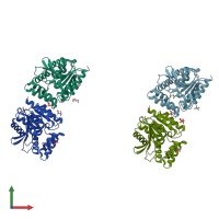
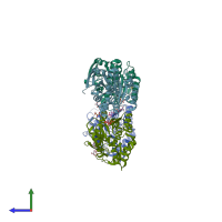
2wj3

X-ray diffraction
2.09Å resolution

CRYSTAL STRUCTURE OF THE COFACTOR-DEVOID 1-H-3-HYDROXY-4- OXOQUINALDINE 2,4-DIOXYGENASE (HOD) FROM ARTHROBACTER NITROGUAJACOLICUS RU61A

Released:

Function and Biology Details

Reaction catalysed:
3-hydroxy-2-methyl-1H-quinolin-4-one + O(2) = N-acetylanthranilate + CO
Biochemical function:
Cellular component:
  • not assigned

Structure analysis Details

Assembly composition:
monomeric (preferred)
PDBe Complex ID:
PDB-CPX-128278 (preferred)
Entry contents:
1 distinct polypeptide molecule
Macromolecule:
1H-3-hydroxy-4-oxoquinaldine 2,4-dioxygenase Chains: A, B, C, D
Molecule details ›
Chains: A, B, C, D
Length: 276 amino acids
Theoretical weight: 31.88 KDa
Source organism: Paenarthrobacter nitroguajacolicus
Expression system: Escherichia coli
UniProt:
  • Canonical: O31266 (Residues: 1-276; Coverage: 100%)
Gene names: ARUE_113p00080, hod, meqE, pAL1.008
Sequence domains: alpha/beta hydrolase fold
Structure domains:

Ligands and Environments

2 bound ligands:
No modified residues

Experiments and Validation Details

Entry percentile scores
X-ray source: DIAMOND BEAMLINE I04
Spacegroup: P212121
Unit cell:
a: 45.17Å b: 167.86Å c: 168.03Å
α: 90° β: 90° γ: 90°
R-values:
R R work R free
0.198 0.196 0.232
Expression system: Escherichia coli