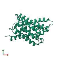
2o9e

X-ray diffraction
2.2Å resolution

Crystal Structure of AqpZ mutant T183C complexed with mercury

Released:
Source organism: Escherichia coli
Primary publication:
Structural basis of aquaporin inhibition by mercury.
J Mol Biol 368 607-17 (2007)
PMID: 17376483

Function and Biology Details

Structure analysis Details

Assembly composition:
homo tetramer (preferred)
Assembly name:
PDBe Complex ID:
PDB-CPX-158108 (preferred)
Entry contents:
1 distinct polypeptide molecule
Macromolecule:
Aquaporin Z Chain: A
Molecule details ›
Chain: A
Length: 234 amino acids
Theoretical weight: 23.92 KDa
Source organism: Escherichia coli
Expression system: Escherichia coli
UniProt:
  • Canonical: P60844 (Residues: 1-231; Coverage: 100%)
Gene names: JW0859, aqpZ, b0875, bniP
Sequence domains: Major intrinsic protein
Structure domains: Glycerol uptake facilitator protein.

Ligands and Environments

1 bound ligand:
No modified residues

Experiments and Validation Details

Entry percentile scores
X-ray source: ALS BEAMLINE 8.3.1
Spacegroup: I4
Unit cell:
a: 91.103Å b: 91.103Å c: 77.944Å
α: 90° β: 90° γ: 90°
R-values:
R R work R free
0.196 0.193 0.243
Expression system: Escherichia coli