2ant

X-ray diffraction
2.6Å resolution

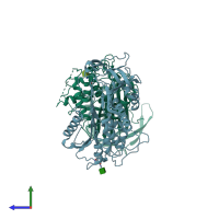
THE 2.6 A STRUCTURE OF ANTITHROMBIN INDICATES A CONFORMATIONAL CHANGE AT THE HEPARIN BINDING SITE

Released:

Function and Biology Details

Structure analysis Details

Assembly composition:
monomeric (preferred)
Assembly name:
PDBe Complex ID:
PDB-CPX-133776 (preferred)
Entry contents:
1 distinct polypeptide molecule
Macromolecule:
Antithrombin-III Chains: I, L
Molecule details ›
Chains: I, L
Length: 432 amino acids
Theoretical weight: 49.1 KDa
Source organism: Homo sapiens
UniProt:
  • Canonical: P01008 (Residues: 33-464; Coverage: 100%)
Gene names: AT3, PRO0309, SERPINC1
Sequence domains: Serpin (serine protease inhibitor)
Structure domains:

Ligands and Environments

1 bound ligand:
No modified residues

Experiments and Validation Details

Entry percentile scores
X-ray source: SRS BEAMLINE PX7.2
Spacegroup: P21
Unit cell:
a: 61.41Å b: 98.31Å c: 90.41Å
α: 90° β: 103.32° γ: 90°
R-values:
R R work R free
0.217 0.217 0.29