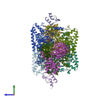
1w88

X-ray diffraction
2.3Å resolution

The crystal structure of pyruvate dehydrogenase E1(D180N,E183Q) bound to the peripheral subunit binding domain of E2

Released:

Function and Biology Details

Structure analysis Details

Assembly composition:
hetero pentamer (preferred)
PDBe Complex ID:
PDB-CPX-146012 (preferred)
Entry contents:
3 distinct polypeptide molecules
Macromolecules (3 distinct):
Pyruvate dehydrogenase E1 component subunit alpha Chains: A, C, E, G
Molecule details ›
Chains: A, C, E, G
Length: 368 amino acids
Theoretical weight: 41.39 KDa
Source organism: Geobacillus stearothermophilus
Expression system: Escherichia coli
UniProt:
  • Canonical: P21873 (Residues: 2-369; Coverage: 100%)
Gene name: pdhA
Sequence domains: Dehydrogenase E1 component
Structure domains: Rossmann fold
Pyruvate dehydrogenase E1 component subunit beta Chains: B, D, F, H
Molecule details ›
Chains: B, D, F, H
Length: 324 amino acids
Theoretical weight: 35.36 KDa
Source organism: Geobacillus stearothermophilus
Expression system: Escherichia coli
UniProt:
  • Canonical: P21874 (Residues: 2-325; Coverage: 100%)
Gene name: pdhB
Sequence domains:
Structure domains:
Dihydrolipoyllysine-residue acetyltransferase component of pyruvate dehydrogenase complex Chains: I, J
Molecule details ›
Chains: I, J
Length: 49 amino acids
Theoretical weight: 5.26 KDa
Source organism: Geobacillus stearothermophilus
Expression system: Escherichia coli
UniProt:
  • Canonical: P11961 (Residues: 123-171; Coverage: 11%)
Gene name: pdhC
Sequence domains: e3 binding domain
Structure domains: E3-binding domain

Ligands and Environments


Cofactor: Ligand TPP 4 x TPP
1 bound ligand:
No modified residues

Experiments and Validation Details

Entry percentile scores
X-ray source: ESRF BEAMLINE ID14-2
Spacegroup: P212121
Unit cell:
a: 92.18Å b: 133.69Å c: 245.61Å
α: 90° β: 90° γ: 90°
R-values:
R R work R free
0.202 0.199 0.262
Expression system: Escherichia coli