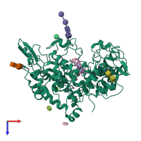
1u67

X-ray diffraction
3.1Å resolution

Crystal Structure of Arachidonic Acid Bound to a Mutant of Prostagladin H Synthase-1 that Forms Predominantly 11-HPETE.

Released:

Function and Biology Details

Reaction catalysed:
Arachidonate + AH(2) + 2 O(2) = prostaglandin H(2) + A + H(2)O
Biochemical function:
Biological process:
Cellular component:
  • not assigned

Structure analysis Details

Assembly composition:
homo dimer (preferred)
PDBe Complex ID:
PDB-CPX-138844 (preferred)
Entry contents:
1 distinct polypeptide molecule
Macromolecules (3 distinct):
Prostaglandin G/H synthase 1 Chain: A
Molecule details ›
Chain: A
Length: 600 amino acids
Theoretical weight: 68.78 KDa
Source organism: Ovis aries
Expression system: Spodoptera frugiperda
UniProt:
  • Canonical: P05979 (Residues: 1-600; Coverage: 100%)
Gene names: COX1, PTGS1
Sequence domains: Animal haem peroxidase
Structure domains:

Ligands and Environments


Cofactor: Ligand COH 1 x COH
Carbohydrate polymer : NEW Components: NAG, NDG
Carbohydrate polymer : NEW Components: NAG, BMA
2 bound ligands:
No modified residues

Experiments and Validation Details

Entry percentile scores
X-ray source: APS BEAMLINE 19-ID
Spacegroup: P6522
Unit cell:
a: 181.879Å b: 181.879Å c: 103.368Å
α: 90° β: 90° γ: 120°
R-values:
R R work R free
0.227 0.238 0.311
Expression system: Spodoptera frugiperda