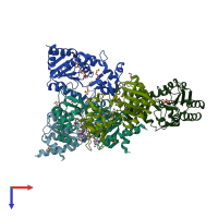
1s7g

X-ray diffraction
2.3Å resolution

Structural Basis for the Mechanism and Regulation of Sir2 Enzymes

Released:
Source organism: Archaeoglobus fulgidus
Primary publication:
Structural basis for the mechanism and regulation of Sir2 enzymes.
Mol Cell 13 639-48 (2004)
PMID: 15023335

Function and Biology Details

Reaction catalysed:
(1a) [protein]-N(6)-acetyl-L-lysine + NAD(+) = [protein]-N(6)-(1,1-(5-adenosylyl-alpha-D-ribose-1,2-di-O-yl)ethyl)-L-lysine + nicotinamide
Biochemical function:
Biological process:
Cellular component:

Structure analysis Details

Assembly composition:
homo pentamer (preferred)
PDBe Complex ID:
PDB-CPX-128216 (preferred)
Entry contents:
1 distinct polypeptide molecule
Macromolecule:
NAD-dependent protein deacylase 2 Chains: A, B, C, D, E
Molecule details ›
Chains: A, B, C, D, E
Length: 253 amino acids
Theoretical weight: 28.54 KDa
Source organism: Archaeoglobus fulgidus
Expression system: Escherichia coli
UniProt:
  • Canonical: O30124 (Residues: 1-253; Coverage: 100%)
Gene names: AF_0112, Sir2Af2, cobB2
Sequence domains: Sir2 family
Structure domains:

Ligands and Environments

Experiments and Validation Details

Entry percentile scores
X-ray source: NSLS BEAMLINE X25
Spacegroup: P21212
Unit cell:
a: 104.85Å b: 181.574Å c: 78.921Å
α: 90° β: 90° γ: 90°
R-values:
R R work R free
0.207 0.207 0.249
Expression system: Escherichia coli