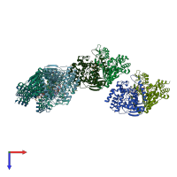
1s20

X-ray diffraction
2.2Å resolution

A novel NAD binding protein revealed by the crystal structure of E. Coli 2,3-diketogulonate reductase (YiaK) NORTHEAST STRUCTURAL GENOMICS CONSORTIUM TARGET ER82

Released:

Function and Biology Details

Reaction catalysed:
3-dehydro-L-gulonate + NAD(P)(+) = (4R,5S)-4,5,6-trihydroxy-2,3-dioxohexanoate + NAD(P)H
Biochemical function:
Biological process:
  • not assigned
Cellular component:

Structure analysis Details

Assembly composition:
homo dimer (preferred)
PDBe Complex ID:
PDB-CPX-153464 (preferred)
Entry contents:
1 distinct polypeptide molecule
Macromolecule:
2,3-diketo-L-gulonate reductase Chains: A, B, C, D, E, F, G, H
Molecule details ›
Chains: A, B, C, D, E, F, G, H
Length: 340 amino acids
Theoretical weight: 38.39 KDa
Source organism: Escherichia coli
Expression system: Escherichia coli
UniProt:
  • Canonical: P37672 (Residues: 1-332; Coverage: 100%)
Gene names: JW3547, b3575, dlgD, yiaK
Sequence domains: Malate/L-lactate dehydrogenase
Structure domains:

Ligands and Environments


Cofactor: Ligand NAD 6 x NAD
1 bound ligand:
1 modified residue:

Experiments and Validation Details

Entry percentile scores
X-ray source: NSLS BEAMLINE X4A
Spacegroup: P1
Unit cell:
a: 75.524Å b: 81.275Å c: 113.215Å
α: 79.55° β: 77.22° γ: 82.01°
R-values:
R R work R free
0.19 0.191 0.25
Expression system: Escherichia coli