1r1p

X-ray diffraction
1.8Å resolution

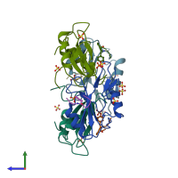
Structural Basis for Differential Recognition of Tyrosine Phosphorylated Sites in the Linker for Activation of T cells (LAT) by the Adaptor Protein Gads

Released:

Function and Biology Details

Biochemical function:
  • not assigned
Biological process:
  • not assigned
Cellular component:
  • not assigned
Structure domain:

Structure analysis Details

Assembly composition:
hetero dimer (preferred)
PDBe Complex ID:
PDB-CPX-131608 (preferred)
Entry contents:
2 distinct polypeptide molecules
Macromolecules (2 distinct):
GRB2-related adaptor protein 2 Chains: A, B, C, D
Molecule details ›
Chains: A, B, C, D
Length: 100 amino acids
Theoretical weight: 11.82 KDa
Source organism: Mus musculus
Expression system: Escherichia coli
UniProt:
  • Canonical: O89100 (Residues: 50-147; Coverage: 30%)
Gene names: Gads, Grap2, Grb2l, Grid, Mona
Sequence domains: SH2 domain
Structure domains: SH2 domain
LAT pY171 peptide Chains: E, F, G, H
Molecule details ›
Chains: E, F, G, H
Length: 7 amino acids
Theoretical weight: 830 Da
Source organism: Mus musculus
Expression system: Not provided

Ligands and Environments

1 bound ligand:
1 modified residue:

Experiments and Validation Details

Entry percentile scores
X-ray source: RIGAKU RU200
Spacegroup: P41212
Unit cell:
a: 90.307Å b: 90.307Å c: 145.959Å
α: 90° β: 90° γ: 90°
R-values:
R R work R free
0.198 0.196 0.241
Expression systems:
  • Escherichia coli
  • Not provided