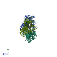
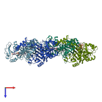
1pqu

X-ray diffraction
1.92Å resolution

Crystal Structure of the H277N Mutant of Aspartate Semialdehyde Dehydrogenase from Haemophilus influenzae Bound with NADP, S-methyl cysteine sulfoxide and cacodylate

Released:
Primary publication:
The role of substrate-binding groups in the mechanism of aspartate-beta-semialdehyde dehydrogenase.
Acta Crystallogr D Biol Crystallogr 60 1388-95 (2004)
PMID: 15272161

Function and Biology Details

Reaction catalysed:
L-aspartate 4-semialdehyde + phosphate + NADP(+) = L-4-aspartyl phosphate + NADPH
Biochemical function:
Cellular component:
  • not assigned

Structure analysis Details

Assembly composition:
homo dimer (preferred)
PDBe Complex ID:
PDB-CPX-155210 (preferred)
Entry contents:
1 distinct polypeptide molecule
Macromolecule:
Aspartate-semialdehyde dehydrogenase Chains: A, B, C, D
Molecule details ›
Chains: A, B, C, D
Length: 371 amino acids
Theoretical weight: 40.56 KDa
Source organism: Haemophilus influenzae Rd KW20
Expression system: Escherichia coli BL21
UniProt:
  • Canonical: P44801 (Residues: 1-371; Coverage: 100%)
Gene names: HI_0646, asd
Sequence domains:
Structure domains:

Ligands and Environments


Cofactor: Ligand NAP 3 x NAP
2 bound ligands:
No modified residues

Experiments and Validation Details

Entry percentile scores
X-ray source: APS BEAMLINE 19-BM
Spacegroup: P21
Unit cell:
a: 72.929Å b: 76.579Å c: 134.097Å
α: 90° β: 92.59° γ: 90°
R-values:
R R work R free
0.184 0.182 0.234
Expression system: Escherichia coli BL21