1out

X-ray diffraction
2.3Å resolution

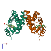
TROUT HEMOGLOBIN I

Released:
Source organism: Oncorhynchus mykiss
Primary publication:
The crystal structures of trout Hb I in the deoxy and carbonmonoxy forms.
J Mol Biol 259 749-60 (1996)
PMID: 8683580

Function and Biology Details

Structure analysis Details

Assembly composition:
hetero tetramer (preferred)
Assembly name:
PDBe Complex ID:
PDB-CPX-135393 (preferred)
Entry contents:
2 distinct polypeptide molecules
Macromolecules (2 distinct):
Hemoglobin subunit alpha-1 Chain: A
Molecule details ›
Chain: A
Length: 143 amino acids
Theoretical weight: 15.11 KDa
Source organism: Oncorhynchus mykiss
Expression system: Not provided
UniProt:
  • Canonical: P02019 (Residues: 1-144; Coverage: 99%)
Gene name: hba1
Sequence domains: Globin
Structure domains: Globins
Hemoglobin subunit beta-1 Chain: B
Molecule details ›
Chain: B
Length: 146 amino acids
Theoretical weight: 15.9 KDa
Source organism: Oncorhynchus mykiss
Expression system: Not provided
UniProt:
  • Canonical: P02142 (Residues: 1-146; Coverage: 100%)
Gene name: hbb1
Sequence domains: Globin
Structure domains: Globins

Ligands and Environments

1 bound ligand:
No modified residues

Experiments and Validation Details

Entry percentile scores
X-ray source: SRS BEAMLINE PX9.6
Spacegroup: P6522
Unit cell:
a: 63.28Å b: 63.28Å c: 312.74Å
α: 90° β: 90° γ: 120°
R-values:
R R work R free
0.162 0.162 0.247
Expression system: Not provided