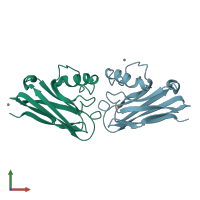
1nwp

X-ray diffraction
1.6Å resolution

CRYSTALLOGRAPHIC STUDY OF AZURIN FROM PSEUDOMONAS PUTIDA

Released:
Source organism: Pseudomonas putida
Primary publication:
Crystallographic study of azurin from Pseudomonas putida.
Acta Crystallogr D Biol Crystallogr 54 253-68 (1998)
PMID: 9761890

Function and Biology Details

Biochemical function:
Biological process:
  • not assigned
Cellular component:

Structure analysis Details

Assembly composition:
homo dimer (preferred)
Assembly name:
PDBe Complex ID:
PDB-CPX-152683 (preferred)
Entry contents:
1 distinct polypeptide molecule
Macromolecule:
Azurin Chains: A, B
Molecule details ›
Chains: A, B
Length: 128 amino acids
Theoretical weight: 13.74 KDa
Source organism: Pseudomonas putida
UniProt:
  • Canonical: P34097 (Residues: 1-128; Coverage: 100%)
Sequence domains: Copper binding proteins, plastocyanin/azurin family
Structure domains: Cupredoxins - blue copper proteins

Ligands and Environments

2 bound ligands:
No modified residues

Experiments and Validation Details

Entry percentile scores
X-ray source: RIGAKU RUH2R
Spacegroup: P21
Unit cell:
a: 40.76Å b: 51.22Å c: 54.96Å
α: 90° β: 103.12° γ: 90°
R-values:
R R work R free
0.181 0.181 0.248