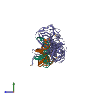
1lwt

X-ray diffraction
3.2Å resolution

Crystal structure of the intein homing endonuclease PI-SceI bound to its substrate DNA (Ca2+ free)

Released:

Function and Biology Details

Reaction catalysed:
ATP + H(2)O + 4 H(+)(Side 1) = ADP + phosphate + 4 H(+)(Side 2)
Biochemical function:
Biological process:
Cellular component:
  • not assigned

Structure analysis Details

Assembly composition:
hetero trimer (preferred)
PDBe Complex ID:
PDB-CPX-114154 (preferred)
Entry contents:
1 distinct polypeptide molecule
2 distinct DNA molecules
Macromolecules (3 distinct):
Endonuclease PI-SceI Chain: A
Molecule details ›
Chain: A
Length: 454 amino acids
Theoretical weight: 51.38 KDa
Source organism: Saccharomyces cerevisiae
Expression system: Escherichia coli
UniProt:
  • Canonical: P17255 (Residues: 284-737; Coverage: 42%)
Gene names: CLS8, D1286, TFP1, VMA1, YDL185W
Sequence domains:
Structure domains:
PI-SceI DNA substrate top strand Chain: B
Molecule details ›
Chain: B
Length: 37 nucleotides
Theoretical weight: 11.6 KDa
Source organism: Saccharomyces cerevisiae
Expression system: Not provided
PI-SceI DNA substrate bottom strand Chain: C
Molecule details ›
Chain: C
Length: 37 nucleotides
Theoretical weight: 11.17 KDa
Source organism: Saccharomyces cerevisiae
Expression system: Not provided

Ligands and Environments

No bound ligands
1 modified residue:

Experiments and Validation Details

Entry percentile scores
X-ray source: APS BEAMLINE 14-BM-C
Spacegroup: P212121
Unit cell:
a: 45.2Å b: 87.4Å c: 214.8Å
α: 90° β: 90° γ: 90°
R-values:
R R work R free
0.248 0.242 0.287
Expression systems:
  • Escherichia coli
  • Not provided