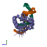
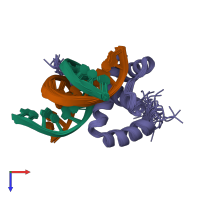
1lfu

Solution NMR

NMR Solution Structure of the Extended PBX Homeodomain Bound to DNA

Released:
Source organism: Mus musculus
Primary publication:
Lock and key binding of the HOX YPWM peptide to the PBX homeodomain.
J Biol Chem 278 1053-8 (2003)
PMID: 12409300

Function and Biology Details

Biochemical function:
Cellular component:
  • not assigned

Structure analysis Details

Assembly composition:
hetero trimer (preferred)
PDBe Complex ID:
PDB-CPX-114138 (preferred)
Entry contents:
1 distinct polypeptide molecule
2 distinct DNA molecules
Macromolecules (3 distinct):
Pre-B-cell leukemia transcription factor 1 Chain: P
Molecule details ›
Chain: P
Length: 82 amino acids
Theoretical weight: 9.58 KDa
Source organism: Mus musculus
Expression system: Escherichia coli
UniProt:
  • Canonical: P41778 (Residues: 233-313; Coverage: 19%)
Gene names: Pbx-1, Pbx1
Sequence domains: Homeobox KN domain
Structure domains: Homeodomain-like
5'-D(*GP*CP*GP*CP*AP*TP*GP*AP*TP*TP*GP*CP*CP*C)-3' Chain: A
Molecule details ›
Chain: A
Length: 14 nucleotides
Theoretical weight: 4.26 KDa
Source organism: Mus musculus
Expression system: Not provided
5'-D(*GP*GP*GP*CP*AP*AP*TP*CP*AP*TP*GP*CP*GP*C)-3' Chain: B
Molecule details ›
Chain: B
Length: 14 nucleotides
Theoretical weight: 4.31 KDa
Source organism: Mus musculus
Expression system: Not provided

Ligands and Environments

No bound ligands
No modified residues

Experiments and Validation Details

Entry percentile scores
Refinement method: dynamical annealing
Expression systems:
  • Escherichia coli
  • Not provided