
-1-PIPERAZINE ETHANESULFONIC ACID</span>;</li> <li class='image_legend_li'>1 copy of <span class='highlight'>water</span>.</li></ul>")
-1-PIPERAZINE ETHANESULFONIC ACID</span>;</li> <li class='image_legend_li'>1 copy of <span class='highlight'>water</span>.</li></ul>")
-1-PIPERAZINE ETHANESULFONIC ACID</span>;</li> <li class='image_legend_li'>1 copy of <span class='highlight'>water</span>.</li></ul>")
Function and Biology Details
Biochemical function:
Biological process:
Cellular component:
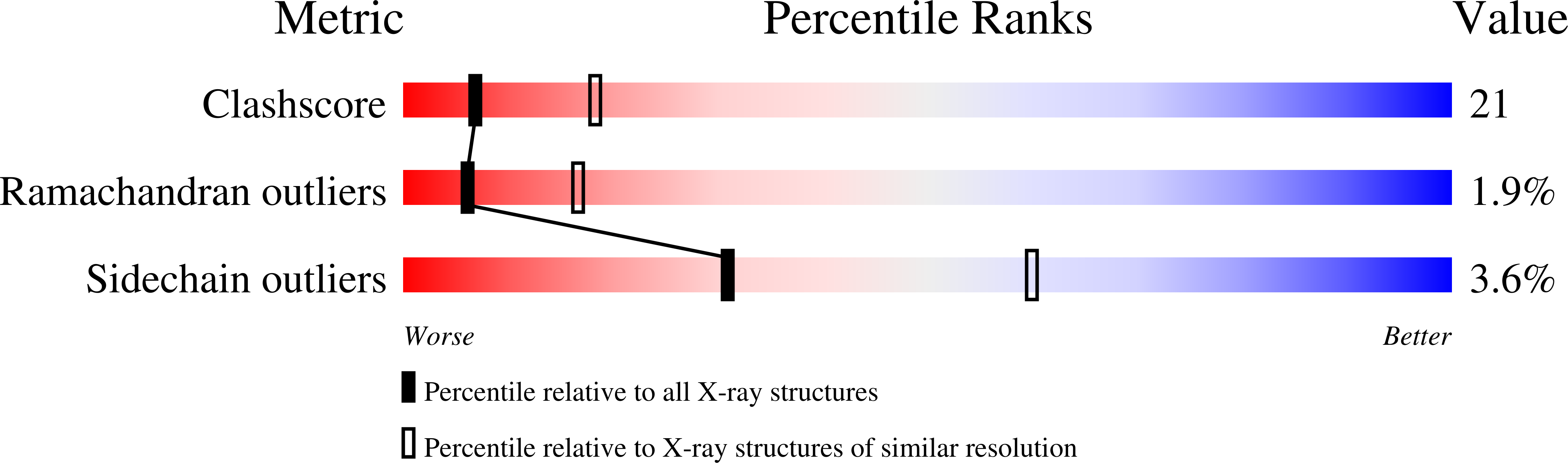
Structure analysis Details
Assemblies composition:
Assembly name:
Acetylcholine binding protein complex (preferred)
PDBe Complex ID:
PDB-CPX-157653 (preferred)
Entry contents:
1 distinct polypeptide molecule
Macromolecule:
Acetylcholine-binding protein
Molecule details ›
Chains: A, B, C, D, E
Length: 217 amino acids
Theoretical weight: 24.69 KDa
Source organism: Lymnaea stagnalis
Expression system: Komagataella pastoris
UniProt:
Structure domains: Neurotransmitter-gated ion-channel ligand-binding domain
Length: 217 amino acids
Theoretical weight: 24.69 KDa
Source organism: Lymnaea stagnalis
Expression system: Komagataella pastoris
UniProt:
- Canonical:
 P58154 (Residues: 14-229; Coverage: 100%)
Structure domains: Neurotransmitter-gated ion-channel ligand-binding domain


