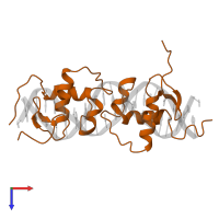
Assemblies
Assembly Name:
Glucocorticoid receptor and DNA
Multimeric state:
hetero tetramer
Accessible surface area:
14973.63 Å2
Buried surface area:
5634.73 Å2
Dissociation area:
1,134.68
Å2
Dissociation energy (ΔGdiss):
8.35
kcal/mol
Dissociation entropy (TΔSdiss):
11.34
kcal/mol
Symmetry number:
1
PDBe Complex ID:
PDB-CPX-113872
Macromolecules
Chains: A, B
Length: 81 amino acids
Theoretical weight: 9.13 KDa
Source organism: Rattus norvegicus
Expression system: Escherichia coli
UniProt:
Pfam: Zinc finger, C4 type (two domains)
InterPro:
CATH: Erythroid Transcription Factor GATA-1, subunit A
SCOP: Nuclear receptor
Length: 81 amino acids
Theoretical weight: 9.13 KDa
Source organism: Rattus norvegicus
Expression system: Escherichia coli
UniProt:
- Canonical:
 P06536 (Residues: 436-514; Coverage: 10%)
Pfam: Zinc finger, C4 type (two domains)
InterPro:
CATH: Erythroid Transcription Factor GATA-1, subunit A
SCOP: Nuclear receptor
Name:
DNA (5'-D(*CP*CP*AP*GP*AP*AP*CP*AP*TP*CP*GP*AP*TP*GP*TP*TP*C P*TP*G)-3')
Representative chains: C, D
Source organism: Rattus norvegicus [10116]
Expression system: Not provided
Length: 19 nucleotides
Theoretical weight: 5.8 KDa
Representative chains: C, D
Source organism: Rattus norvegicus [10116]
Expression system: Not provided
Length: 19 nucleotides
Theoretical weight: 5.8 KDa


