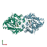
1ez2

X-ray diffraction
1.9Å resolution

THREE-DIMENSIONAL STRUCTURE OF THE ZINC-CONTAINING PHOSPHOTRIESTERASE WITH BOUND SUBSTRATE ANALOG DIISOPROPYLMETHYL PHOSPHONATE.

Released:
Source organism: Brevundimonas diminuta
Primary publication:
The binding of substrate analogs to phosphotriesterase.
J Biol Chem 275 30556-60 (2000)
PMID: 10871616

Function and Biology Details

Reaction catalysed:
An aryl dialkyl phosphate + H(2)O = dialkyl phosphate + an aryl alcohol
Biochemical function:
Biological process:
Cellular component:

Structure analysis Details

Assembly composition:
homo dimer (preferred)
Assembly name:
PDBe Complex ID:
PDB-CPX-141251 (preferred)
Entry contents:
1 distinct polypeptide molecule
Macromolecule:
Parathion hydrolase Chains: A, B
Molecule details ›
Chains: A, B
Length: 331 amino acids
Theoretical weight: 35.96 KDa
Source organism: Brevundimonas diminuta
Expression system: Escherichia coli
UniProt:
  • Canonical: P0A434 (Residues: 35-365; Coverage: 99%)
Gene name: opd
Sequence domains: Phosphotriesterase family
Structure domains: Metal-dependent hydrolases

Ligands and Environments

2 bound ligands:
1 modified residue:

Experiments and Validation Details

Entry percentile scores
X-ray source: RIGAKU RU200
Spacegroup: C2
Unit cell:
a: 129.4Å b: 91Å c: 69.2Å
α: 90° β: 91.6° γ: 90°
R-values:
R R work R free
0.183 not available not available
Expression system: Escherichia coli