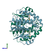
1dug

X-ray diffraction
1.8Å resolution

STRUCTURE OF THE FIBRINOGEN G CHAIN INTEGRIN BINDING AND FACTOR XIIIA CROSSLINKING SITES OBTAINED THROUGH CARRIER PROTEIN DRIVEN CRYSTALLIZATION

Released:

Function and Biology Details

Structure analysis Details

Assembly composition:
homo dimer (preferred)
PDBe Complex ID:
PDB-CPX-136035 (preferred)
Entry contents:
1 distinct polypeptide molecule
Macromolecule:
Fibrinogen gamma chain; Glutathione S-transferase class-mu 26 kDa isozyme Chains: A, B
Molecule details ›
Chains: A, B
Length: 234 amino acids
Theoretical weight: 27.13 KDa
Source organisms: Expression system: Escherichia coli
UniProt:
  • Canonical: P08515 (Residues: 2-218; Coverage: 100%)
  • Canonical: P02679 (Residues: 424-433; Coverage: 2%)
Gene names: FGG, PRO2061
Sequence domains:
Structure domains:

Ligands and Environments


Cofactor: Ligand GSH 2 x GSH
No bound ligands
No modified residues

Experiments and Validation Details

Entry percentile scores
X-ray source: NSLS BEAMLINE X4A
Spacegroup: P41212
Unit cell:
a: 105.78Å b: 105.78Å c: 137.23Å
α: 90° β: 90° γ: 90°
R-values:
R R work R free
0.185 0.185 0.226
Expression system: Escherichia coli