


Function and Biology Details
Reaction catalysed:
ATP + a [protein]-L-tyrosine = ADP + a [protein]-L-tyrosine phosphate
Biochemical function:
- not assigned
Biological process:
- not assigned
Cellular component:
- not assigned
Sequence domains:
Structure domain:
Structure analysis Details
Assembly composition:
homo tetramer (preferred)
Assembly name:
EphB2/CTF2 (preferred)
PDBe Complex ID:
PDB-CPX-151408 (preferred)
Entry contents:
1 distinct polypeptide molecule
Macromolecule:
EphB2/CTF2
Molecule details ›
Chains: A, B, C, D, E, F, G, H
Length: 82 amino acids
Theoretical weight: 9.4 KDa
Source organism: Homo sapiens
Expression system: Escherichia coli BL21
UniProt:
Sequence domains: SAM domain (Sterile alpha motif)
Structure domains: Transcription Factor, Ets-1
Length: 82 amino acids
Theoretical weight: 9.4 KDa
Source organism: Homo sapiens
Expression system: Escherichia coli BL21
UniProt:
- Canonical:
 P29323 (Residues: 905-985; Coverage: 8%)
Sequence domains: SAM domain (Sterile alpha motif)
Structure domains: Transcription Factor, Ets-1
Ligands and Environments
No bound ligands
No modified residues
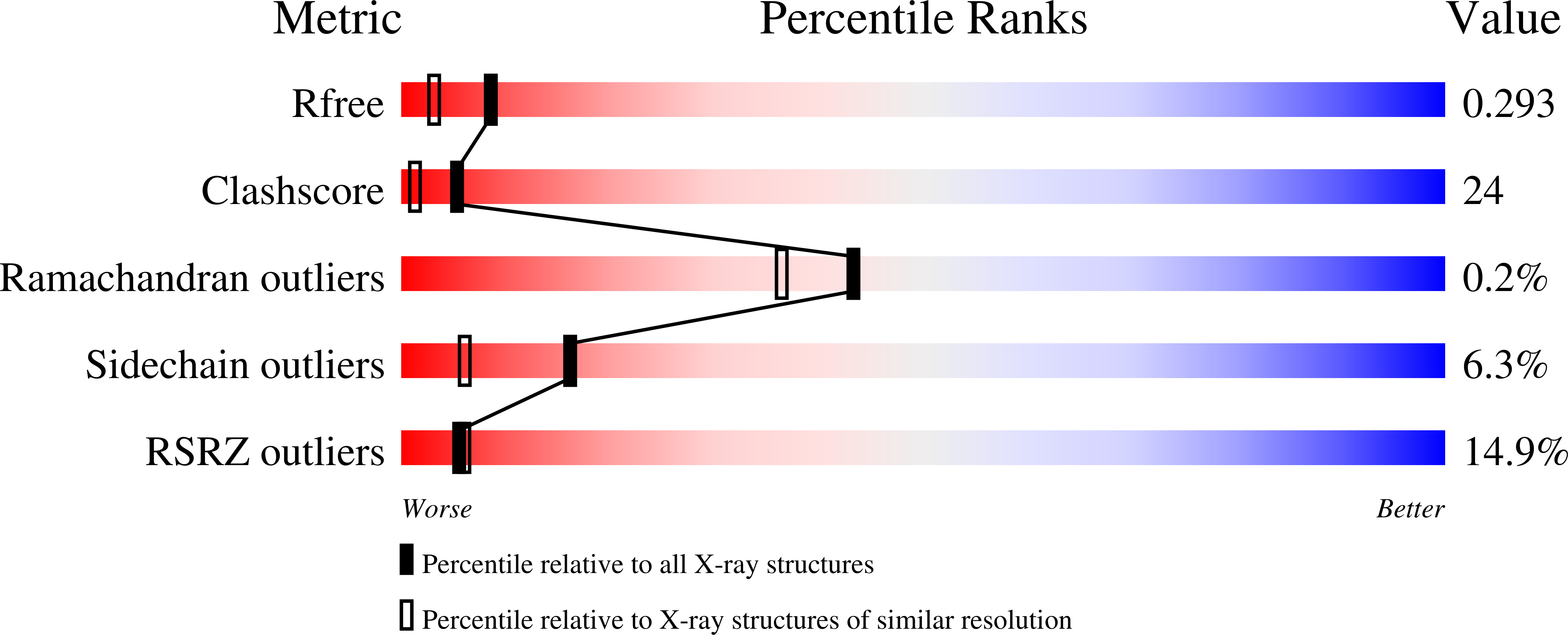
Experiments and Validation Details

X-ray source:
ALS BEAMLINE 5.0.2
Spacegroup:
P41
Expression system: Escherichia coli BL21