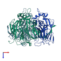
1mzz

X-ray diffraction
2Å resolution

Crystal Structure of Mutant (M182T)of Nitrite Reductase

Released:
Source organism: Cereibacter sphaeroides
Entry authors: Guo H, Olesen K, Xue Y, Shapliegh J, Sjolin L

Function and Biology Details

Reaction catalysed:
Nitric oxide + H(2)O + ferricytochrome c = nitrite + ferrocytochrome c + 2 H(+)
Biological process:
Cellular component:

Structure analysis Details

Assembly composition:
homo trimer (preferred)
PDBe Complex ID:
PDB-CPX-176049 (preferred)
Entry contents:
1 distinct polypeptide molecule
Macromolecule:
Copper-containing nitrite reductase Chains: A, B, C
Molecule details ›
Chains: A, B, C
Length: 334 amino acids
Theoretical weight: 36.42 KDa
Source organism: Cereibacter sphaeroides
Expression system: Escherichia coli
UniProt:
  • Canonical: Q53239 (Residues: 39-371; Coverage: 97%)
Gene names: Rsph17025_1595, nirK
Sequence domains:
Structure domains: Cupredoxins - blue copper proteins

Ligands and Environments

1 bound ligand:
No modified residues

Experiments and Validation Details

Entry percentile scores
X-ray source: RIGAKU RU300
Spacegroup: P212121
Unit cell:
a: 92.495Å b: 123.729Å c: 130.173Å
α: 90° β: 90° γ: 90°
R-values:
R R work R free
0.17 0.17 0.218
Expression system: Escherichia coli