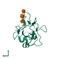
1iko

X-ray diffraction
1.92Å resolution

CRYSTAL STRUCTURE OF THE MURINE EPHRIN-B2 ECTODOMAIN

Released:
Source organism: Mus musculus
Primary publication:
Crystal structure of an ephrin ectodomain.
Dev Cell 1 83-92 (2001)
PMID: 11703926

Function and Biology Details

Structure analysis Details

Assembly composition:
monomeric (preferred)
Assembly name:
PDBe Complex ID:
PDB-CPX-156645 (preferred)
Entry contents:
1 distinct polypeptide molecule
Macromolecules (2 distinct):
Ephrin-B2 Chain: P
Molecule details ›
Chain: P
Length: 178 amino acids
Theoretical weight: 19.9 KDa
Source organism: Mus musculus
Expression system: Komagataella pastoris
UniProt:
  • Canonical: P52800 (Residues: 30-207; Coverage: 58%)
Gene names: Efnb2, Elf2, Epl5, Eplg5, Htkl, Lerk5
Sequence domains: Ephrin
Structure domains: Cupredoxins - blue copper proteins

Ligands and Environments

Carbohydrate polymer : NEW Components: NAG, MAN
No bound ligands
No modified residues

Experiments and Validation Details

Entry percentile scores
X-ray source: APS BEAMLINE 19-ID
Spacegroup: P6522
Unit cell:
a: 59.283Å b: 59.283Å c: 168.868Å
α: 90° β: 90° γ: 120°
R-values:
R R work R free
0.216 0.216 0.249
Expression system: Komagataella pastoris